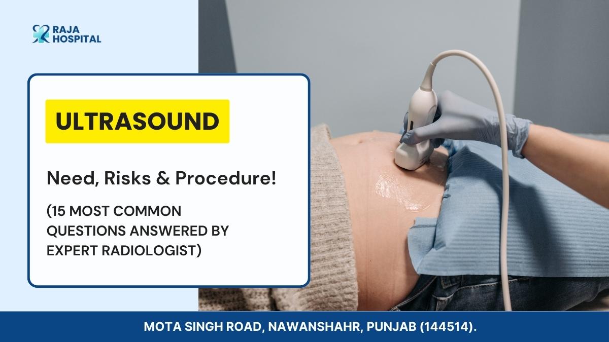
Going for Ultrasound? 15 Myths Busted by Expert Radiologist!
Reviewed By Dr. Amarinder Singh Saini (MBBS, MD Radio Diagnosis) on 15th July 2023.
The visualizing technique of diagnostic ultrasonography, also called sonography or diagnostic medical sonography, utilises sound waves to create pictures of the internal organs and other components in your body.
Several illnesses and ailments may be diagnosed, and therapy can be planned using the photos. While specific ultrasound exams require inserting a tiny ultrasound machine into the human body, most general ultrasound use ultrasound equipment outside your body.
What is Ultrasound? Why Do You Need One?

An ultrasound is generally a medical examination that records real-time pictures of your body’s interior using high-frequency sound propagation. It is sometimes referred to as sonography.
The technology is comparable to sonar along with radar, which aids in the military’s ability to find ships & airplanes. Without having to perform a cut, an ultrasound also be used enables a physician to spot concerns with organs, arteries, and tissues.
In contrast to other visualizing methods, ultrasonography doesn’t employ radiation. It’s hence the procedure for observing a growing foetus throughout pregnancy.
The majority of people connect ultrasounds with maternity. A pregnant woman may see her unborn child for the first time through these ultrasound device. The procedure, however, has a wide range of different applications.
If you have discomfort, swelling, or additional signs that call for a look at the organs of your body, your doctor may recommend an ultrasound. An ultrasound test can show the following:
- blood vessels
- bladder
- uterus
- brain (in infants)
- eyes
- testicles
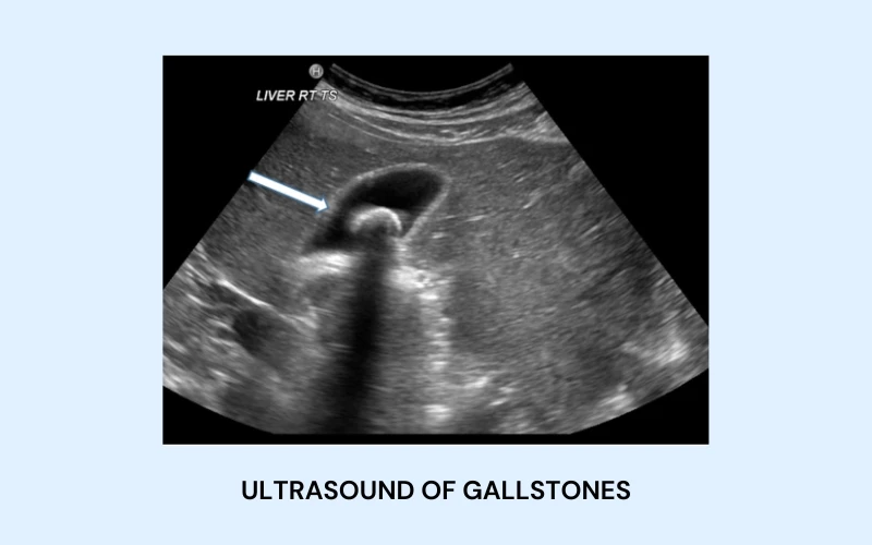
- gallbladder
- kidneys
- liver
- thyroid
- ovaries
- spleen
- pancreas
Using a medical ultrasound also directs surgeon motions during specific medical operations, including biopsies, is also beneficial.
How to Prepare for an Ultrasound Test?
The doctors will confirm your identity and perform the test you ordered. Depending on the kind of ultrasound technique your doctor has recommended, you’ll need to prepare for this exam. Some preparations involve consuming a glass of drinking water for the examination to get better results. Your healthcare provider will provide instructions.
You won’t be allowed to eat or drink after midnight the evening prior to the biopsy if you are having one. Your doctor will provide instructions.
Clothing and Jewelry?
Dress comfortably during your ultrasound visit. During your Ultrasound, you could be requested to take off your jewellery. Therefore, leaving any accessories or jewellery at home is an excellent decision.
Food?
After midnight, avoid eating any food, either solid or liquid. One can, however, mix some liquid along with your medication. Avoid chewing gum since doing so will cause you to swallow air, which might degrade the visuals.
Water?
It’s acceptable to drink water and take medicine. For female patients having an Ultrasound pelvic as well, preferably drink thirty-two ounces of water approximately an hour before the scan. As long as you continue drinking water, you can use the loo.
Risks Involved in Ultrasound Scan:

Low-power ultrasound waves of sound are used during the safe diagnostic ultrasonography process. No risks are noted.
Although an excellent instrument, Ultrasound produces has certain drawbacks. Because it uses high-frequency sound waves which don’t propagate effectively through air or bone, ultrasonography is ineffective at visualizing bodily areas like the brain and lungs that contain gas or are covered by bone.
Additionally, items may be invisible to Ultrasound deep inside the body. To assess these regions, your doctor may request further medical imaging test procedures, including magnetic resonance imaging (MRI) or X-rays.
How Does an Ultrasound Report Look?
Red and blue shades address the growth of blood. Red and blue are used for addressing blood flowing beyond the test, whereas blue is used to address blood flowing onto the test.
If the screen displays a mixture of blue and red, it may depict a circular stream, a logical flow, or a disruption. A range of blue and red hues represents speed. Lighter colours show incredible speed, while darker tones indicate extraordinary slowness.
For example, if an ultrasound consists of a uterus, the tissues are laid out at the peak. As the image descends towards the bottom of the display, the uterine lining & amniotic fluid are seen alongside the deeper tissue. When compared to the highest portion of the uterus plus the tissue higher than it, the peak of a foetal ultrasound resembles bundles of thick tissue. Immediately underneath this, there is a primarily black area that is made up of amniotic fluid.
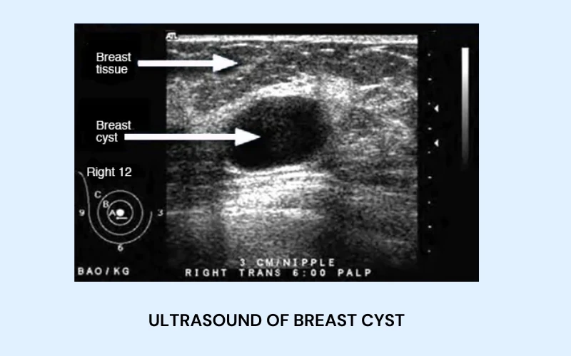
Ultrasound of Breast Cyst
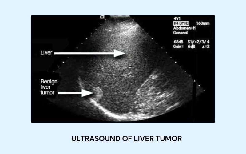
Ultrasound of Liver Tumor
Ultrasound of Gallstones
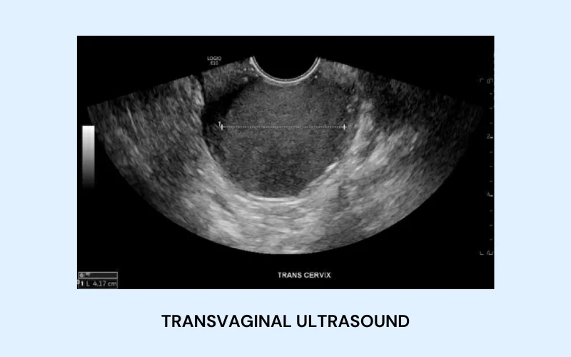
Transvaginal Ultrasound
15 Most Common Questions Regarding Ultrasound Scans:
Here are the most common questions that patients generally ask us. If you have anymore concerns, please feel free to contact our team.
Do frequent ultrasound scans have side effects?
Ultrasound scan is a safe modality , which uses sound to create pictures . It does no harm.
That is why ultrasound scans are safely used in pregnancy . They do not harm the fetus. It has also been proven in various studies.
Transvaginal scan is risky and painful?
Transvaginal scans provide very high resolution images of uterus ,other pelvic organs. It can be safely used to assess pelvic problems in women.
Why can’t I pee before ultrasound scan of abdomen?
Full bladder gives the doctor a good window to check your pelvic organs. If the bladder is not full the pelvic organs are not completely visible, which can lead to wrong or incomplete reporting of disease.
Ultrasound scan done anywhere should give same result!
Ultrasound scan is user dependent and highly skilled procedure . It largely depends on the doctor performing the scan. So the experience of the doctor and quality of ultrasound machine matter a lot.
Will I need to do anything to prepare for the test?
Before a scan of the unborn child or your pelvic region, it may be necessary to consume water and wait to use the loo until after the examination.
A scan should be scheduled many hours before consuming food or drink. Before examining your digestive system, which may include the liver plus gallbladder, this might be required.
What happens during an Ultrasound?
The ultrasound doctor will ask you to lay flat on an examination table. The examiner will treat the outer layer of skin of the region being checked with gel. This makes it simpler to manoeuvre the slick, hand-held transducer. Pictures are produced by the transducer using sound waves.
Do ultrasounds Cause Autism?
There is no link between the quantity or length of prenatal ultrasounds with a child’s subsequent autism diagnosis.
Can Ultrasound detect breast cancer?
In thick breast tissue, Ultrasound is a beneficial diagnostic tool for finding breast tumours that mammography frequently misses.
Are Ultrasound and CT Scans the Same?
Images of bone structures do not frequently need the use of ultrasounds. Contrarily, CT (computerized tomography) scans may deliver in-depth pictures of blood arteries, bones, and even soft tissues. Ultrasounds are frequently utilised in the prenatal treatment and other operations like gallstones or stones from kidney extraction.
Which Ultrasound is Used for Kidney Stones?
Abdominal and pelvic ultrasounds: These tests employ sound waves to produce images of the kidneys and even the bladder, and they can detect obstructions in the flow of urine and assist in identifying stones.
Can Ultrasound help in detecting cancer or tumours?
Doctors can use an ultrasound to check for tumours in parts of the human body that are difficult to see on X-rays. Physicians frequently use this technique to direct a needle throughout a biopsy. Many ultrasounds do not need any prior planning and are often rapid.
How is Ultrasound used in monitoring the development of a fetus during pregnancy?
First trimester: During the initial three weeks of the pregnancy, an ultrasound is utilised to confirm the number of embryos, determine the gestational age, and determine the expected arrival of the baby.
Can Ultrasound detect abnormalities in the organs of the abdomen?
It is employed to examine the liver, the gallbladder, the spleen, the pancreas, and the kidneys, among other abdominal organs. Ultrasound may also examine the circulatory veins connecting to several organs, including the aorta & inferior vena cava.
Are there any factors that can affect the quality of an Ultrasound image?
Fat and air are the two primary elements that interfere with ultrasound quality. Experimental evidence has shown that resolution declines as overlapping fat thickness increases.
What should I eat before an Ultrasound?

A good, light supper is often advised before having an ultrasound. It involves avoiding foods heavy in sugar or saturated fat and anything that can induce indigestion or irritation. Instead, go for fresh produce, lean protein, or whole-wheat bread.
What is the cost of an Ultrasound test?
An ultrasound typically costs between Rs. 600 and Rs. 1500.
Is the Ultrasound done on an empty stomach?
You must fast for Eight to twelve hours before an abdominal ultrasound. We refer to this called fasting. Fasting aids in preventing gas accumulation in the abdominal region, which might influence the outcomes.
Do I need to drink water before Ultrasound?
A full bladder will move the abdomen forward and push the colon out of its position, allowing you to see the womb, cervix, ovaries, baby, and placenta more clearly. If your bladder is full, obtaining the finest pictures and ruling out any issues will be easier.
How long is the Ultrasound process?
The majority of ultrasound examinations continue for 15 to 45 minutes. These are often carried out by a physician, radiographer, or sonographer at the hospital’s radiology laboratory.
How long do Ultrasound results take?
The entire ultrasound examination takes 30 minutes. You’ll often get the answer in less than 24 hours.
Can Ultrasound show infection?
This demonstrated that Ultrasound is an even more accurate diagnostic tool than a clinical exam alone for the examination of infections of the skin and soft tissues, improving our capacity to identify an abscess cavity or, more profoundly, disease.
Can Ultrasound detect fatty liver?
Ultrasonography is a reliable method for spotting fatty liver. It has histology-like specificity and sensitivity when identifying moderate to severe fatty liver.
What is the difference between 2D, 3D, and 4D Ultrasounds?
There are different type of ultrasounds out there. Compared to the flat, two-dimensional, black-and-white pictures utilised in OB/GYN, 2D Ultrasound offers superior image quality. The 3D ultrasound picture in three dimensions is orange and golden. The live feed of the 3D picture is 4D Ultrasound.
What are the benefits of using Ultrasound over other imaging techniques like X-rays or CT scans?
Real-time ultrasound visualization allows for the display of blood flowing via blood vessels and the motion of the body’s interior organs. Unlike X-ray imaging, ultrasonic imaging does not expose patients to ionising radiation.
How is Usg used in monitoring development of fetus during pregnancy?
It is the safest test to perform during pregnancy as it used sound waves and does not harm the baby. Even repeated Usg has no bad effect on baby. Ultrasound is conducted in all trimesters of pregnancy to detect and structural abnormalities in the baby, placenta, cervix and amount of water (liquor) around the baby and blood flow from mother to baby.
What is Ultrasound Guided Biopsy.
When biopsy are taken with the help of ultrasound guidance. It has better results as the abnormal organ is focused and biopsy taken from the required area, leading to better results and reduced re dos.
Is there a special Ultrasound for heart-related issues?
An “echo” scan examines the heart and adjacent blood vessels. It’s a form of ultrasonic scan that uses a tiny probe to transmit high-frequency sound waves that produce echoes when they reverberate off various bodily organs.
Final Thoughts
In summary, the acoustic impedances of diverse materials significantly emphasise ultrasound technology. The sound waves that bounce back to the transducer’s detector are transformed into graphing and a sharp 2D picture. Without undergoing surgeries or other treatments, it is now feasible to analyse the structure of human beings via this kind of technology.